
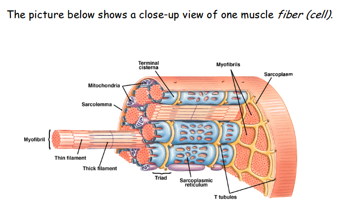
The muscles contract and contain energy by the sodium potassium pump (Na+ K+ Pump). At the neuromuscular junction ACh is released by synaptic terminal and binds to receptors on sarcolemma. Resulting change in membrane potential leads to production of an action potential that spreads across the entire surface of muscle fiber and along the T-tubules. Sarcoplasmic reticulum releases stored calcium ions, increasing calcium concentration in the sarcoplasm. Calcium ions bind to troponin, resulting in movement of tropomyosin and exposure of active sites on the thin filaments. Crossbridge form when myosin heads bind to active sites. Contraction begins as repeated cycles of crossbrigde binding, pivoting, and detachment occur, powered by the breakdown of ATP. These events produce filament sliding, and muscle fiber shortens. Action potential generation ceases ACh is broken down by AChE. The sarcolplasmic reticulum reabsorbs calcium ions in the sarcoplasm declines. When calcium ion concentration approach normal resting levels, the troponin and tropomyosin molecules return to their normal positions. This recovers the active sites. With the active sites no longer accessible, crossbridge formation cannot occur. Without crossbridge interactions, further sliding cannot take place, and the contraction ends. Muscle relaxtion occurs, and the muscle returns passively to its resting length.


No comments:
Post a Comment